As a radiologist, i think that the largest mistake is to believe that all X-rays are of good quality… not just the image quality itself, it’s more about the deviations from standard projection, low occurence of pathology mimicking normal variants and so on.
Just read your Article on Medium and liked it very much, but it seems that the preprocessing is kind of missing… some x-rays are scaled down for example, way to much blank spaces around the images themselves.
Hi @Sandorkonya,
As written in my medium post, I applied Data Augmentation (DA) as following:
horizontal flip and max rotate of 30° without wrap.
default fastai transformations as max_zoom=1.1 and max_lighting=0.2 through the get_transforms() fastai function.
I would enjoy apply more accurate preprocessing of the x-rays in DA. Could you suggest some and give the corresponding fastai code? Many thanks in advance.
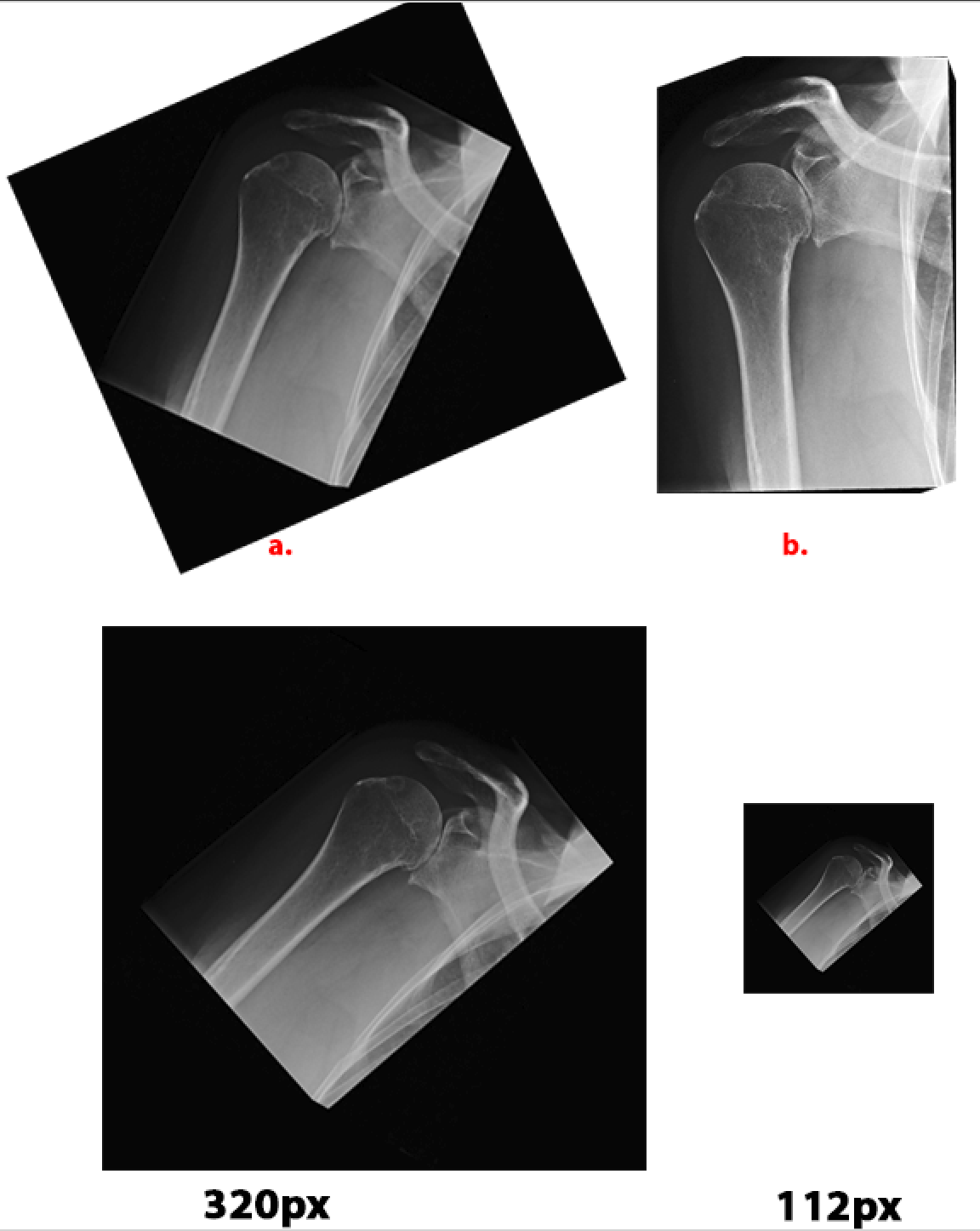
instead of a. the ideal case would be to take b. (cropped, magnified). It would (probably) normalize the image features.
The original would look with 1.1 magnification and a 30° rotation resampled to 320px and 112px above The “black area” of the frame is huge, the image features are very small.
I think an object-detection approach could be utilized, where one trains an object detection model on different x-ray categories (with well captured/croped ) and it “simply” crops and aligns the region from the original x-ray if it is rotated initially or there is to thick frame around it.